Abstract
Erythropoiesis requires a substantial amount of iron, and both congenital and acquired conditions associated with iron overload can lead to early erythroid death. X-linked sideroblastic anemia (XLSA), most common form of congenital sideroblastic anemia, represents this disorder. In XLSA, germline mutations in the erythroid-specific 5-aminolevulinate synthase (ALAS2) gene cause impaired heme biosynthesis and mitochondrial iron overload. Thus, erythroblasts in patients with XLSA undergo premature death; however, the pathological mechanism behind this is yet to be elucidated. We recently established cellular XLSA models using human umbilical cord blood-derived erythroid progenitor (HUDEP)-2 cells (Ono et al. Sci Rep 2022). Remarkably, XLSA clones (ALAS2 R170L and R170H) had notably low heme concentration and high transcription factor BACH1 due to decreased heme-mediated degradation. Consequently, genes repressed by BACH1 had reduced expression. Some downregulated genes were associated with ferroptosis inhibitory mechanisms, such as glutathione synthesis and iron metabolism. In XLSA clones, intracellular glutathione levels were significantly reduced, whereas lipid peroxidation levels were elevated. These findings suggest that BACH1 promotes XLSA clones prone to ferroptosis by transcriptionally regulating gene expression. However, ferroptosis occurs only when XLSA clones are treated with a ferroptosis inducer, erastin. Some mechanisms are assumed to inhibit ferroptosis during erythroid differentiation in XLSA. Here, we focused on the master transcription factor GATA1 that regulates erythroid differentiation and survival, demonstrating how XLSA clones inhibit ferroptosis.
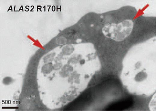
After erythroid differentiation, XLSA clones showed a decreased proportion of mature erythroblasts compared to the wild type, indicating an impaired differentiation in XLSA clones. Heme plays an essential role in erythroid differentiation, and heme deficiency can impair XLSA clone differentiation. Intriguingly, we observed that GATA1 protein expression, induced during erythropoiesis, was notably higher in XLSA clones than in the wild type. Mass spectrometry-based proteomic analysis revealed that XLSA clones globally upregulated stress-inducible proteins such as HSPA9, whereas caspase-3 was inactivated. These findings were consistent with previous description stating that chaperon proteins, which are induced by oxidative stress, prevent caspase-mediated GATA1 proteolysis (Ribeil et al. Nature 2007). Next, we investigated the autophagic activity of XLSA clones, because GATA1 regulates gene expression associated with autophagy mechanisms, which are important for terminal erythropoiesis. GATA1 has been known to directly activate the transcription of autophagy-related genes, such as BNIP3L, MAP1LC3B, and GABRAP, and indirectly regulate autophagy by inducing FOXO3. Despite GATA1 accumulation, XLSA clones showed lower gene and protein expressions. Flow cytometric analysis using Cyto-ID showed that XLSA clones were associated with impaired autophagy, and electron microscopy detected the accumulation of large autophagolysosomes, which were not properly expelled (Figure, arrows). Proteins interacting with GATA1 were compared between XLSA clones and wild type, and several proteins alternatively interacting with GATA1 were identified. The list of proteins included histones and transcription factors, suggesting that the protein binding in the GATA1 complex is altered in XLSA clones due to low heme and reduced the gene expression. Autophagy plays a key role in inhibiting ferroptosis by NCOA4-mediated turnover of ferritin, known as ferritinophagy, and NCOA4 was found to be induced during erythroid differentiation, whereas its expression was suppressed in XLSA clones compared to the wild type. XLSA clones showed increased ferritin protein accumulation presumably due to the low activity of ferritinophagy.
Therefore, our findings suggest that heme deficiency due to ALAS2 mutation impairs autophagy and prevents ferroptosis in XLSA, accounting for the intact cell viability of XLSA clones despite the increased ferroptosis susceptibility. Thus causative factors for the destruction of this ferroptosis inhibitory mechanism in patients with XLSA should be examined.
Disclosures
Onodera:Astellas: Honoraria; Janssen: Honoraria; Otsuka: Honoraria; Nippon shinyaku: Honoraria; BMS: Research Funding; Celgene: Research Funding; Agios: Research Funding; Abbvie: Honoraria; Novartis: Honoraria. Fukuhara:Celgene: Honoraria, Research Funding; Ono pharma: Honoraria; Nippon Shinyaku: Honoraria; Kyowa kirin: Honoraria; Janssen: Honoraria; Eisai: Honoraria; Dainippon Sumitomo: Honoraria; Incyte: Research Funding; Genmab: Research Funding; BMS: Honoraria, Research Funding; Chugai pharma: Honoraria, Research Funding; Bayer: Research Funding; Novartis: Consultancy, Honoraria; HUYA: Consultancy; Eli Lilly: Consultancy; AstraZeneca: Consultancy, Honoraria; Abbvie: Consultancy; Sanofi: Honoraria; Symbio: Honoraria; Takeda: Honoraria. Onishi:Meiji Seika: Honoraria; Chugai: Honoraria; Amgen: Honoraria; Symbio: Honoraria; Janssen: Honoraria, Research Funding; Kyowa Kirin: Honoraria; BMS: Honoraria; Abbvie: Honoraria; Takara Bio: Research Funding; Pfizer: Honoraria, Research Funding; Novartis: Honoraria, Research Funding; Astellas: Honoraria; MSD: Honoraria. Yokoyama:Astellas: Honoraria; BMS: Honoraria; Janssen: Honoraria; Sanofi: Honoraria. Harigae:Abbvie: Honoraria, Membership on an entity's Board of Directors or advisory committees; Sanofi: Honoraria, Membership on an entity's Board of Directors or advisory committees; Janssen: Honoraria, Membership on an entity's Board of Directors or advisory committees; Novartis: Honoraria; Chugai: Honoraria; Symbio: Honoraria.
Author notes
Asterisk with author names denotes non-ASH members.


